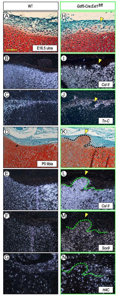
Fig. 2.
Gene expression is altered along mutant chondro-perichondrial border in vivo. (A–G) WT long bone sections at E16.5 and PO (A and D) displaying typical expression patterns of the cartilage markers collagen IIB (B,E) and Sox9 (F) and the perichondrial marker tenascin-C (C). Proliferative H4C-positive cells are present in both perichondrium and cartilage (G). (H–N) Sections from Gdf5-Cre; Ext1fl/fl mutant limbs (H and K) show that the ectopic cartilage within perichondrium expresses collagen IIB (I and L) and Sox9 (M) but not tenascin-C (J) and contains, and is surrounded by, H4C-positive cells (N). Arrowheads indicate the location of ectopic cartilage. Scale bar, 100 μm.

