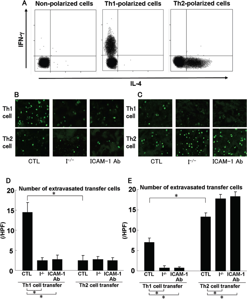
Figure 7.
Cytokine profile of in vitro-generated polarized Th1 or Th2 cell populations (A). Polarized Th1 and Th2 cells were double-stained for intracellular IFN-γ and IL-4 after activation with anti-CD3 and anti-CD28 mAb and were analyzed by flow cytometry. C-AM-labeled polarized Th1 or Th2 cells were injected into wild type mice treated with control Ab (CTL), ICAM-1−/− mice (I−/−), and wild type mice treated with anti-ICAM-1 mAb (ICAM-1 Ab) at 1 hour before DNFB (B, D) or FITC (C, E) elicitation. Mice were sacrificed 24 hours after elicitation. Cell infiltration in the skin was scanned by confocal laser microscopy (B, C). The number of extravasated transfer cells was counted in 10 random grids under magnification of high-power fields (HPF, ×400) and averaged (D, E). Each histogram shows the mean (± SEM) results obtained from 5 mice of each group. *p<0.05.

