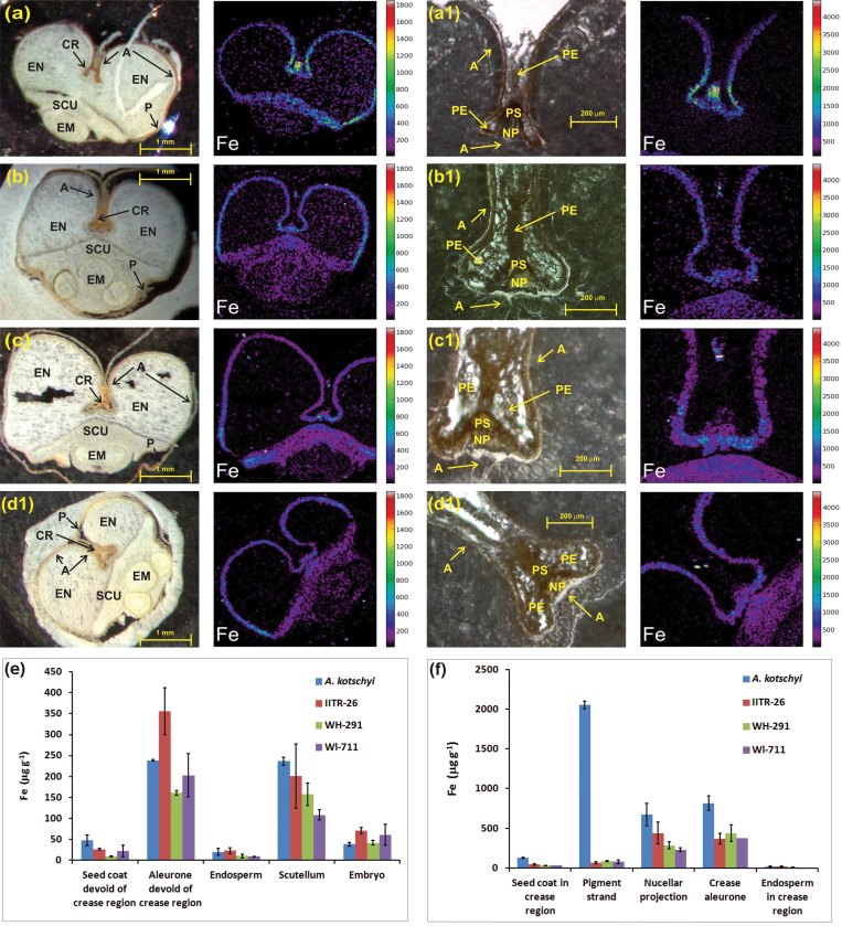
Fig. 2.
(A–D) Micro-PIXE images showing iron localization in cross-section of whole grain (left) and crease tissues (right) of A. kotschyi acc. 3790 (A), T. aestivum L. IITR26 (B), T. aestivum Cv. WH291 (C), and T. aestivum Cv. WL711 (D). (E, F) Iron accumulation patterns based on iron concentration in grain (E) and crease tissues (F) of the four genotypes. Values are mean ± SE μg (g dryweight)–1 from selected tissues (Fig. S2) of two independently measured cuttings. The aleurone and seed coat in E are devoid of crease region. The endosperm in F corresponds only to the region around the crease. A, aleurone; CR, crease; EM, embryo; EN, endosperm; NP, nucellar projection; P, peripheral tissues (pericarp and seed coat); PE, pericarp; PS, pigment (vascular) strand; SCU, scutellum.

