Fig. 2.
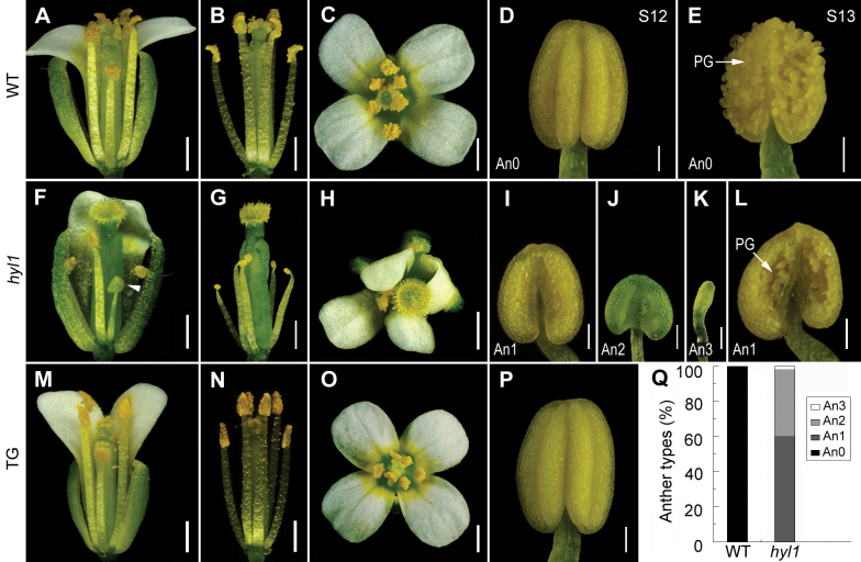
Stamen phenotype of hyl1 mutants. (A–E) The wild-type. (A) Side view of the opened flower. (B) Side view of the same flower as in (A) with all sepals and petals cut off to show the stamens and pistil. (C) Overview of the opened flower. (D) An anther at late stage 12 showing two inner and two outer microsporangia in the adaxial area. (E) An anther with released pollen at stage 13. (F–L) hyl1 mutants. (F) An opened flower with one sepal and two petals cut off to show the stamen defects. (G) A flower with all sepals and petals cut off to show the stamens and pistil. (H) Overview of the opened flower. (I–K) Anthers of An1 (I) at stage 12, An2 (J), and An3 (K). (L) An1 anther at stage 13. (M–P) Flowers and stamens of a hyl1 plant transgenic for HYL1. (Q) Percentages of the three types of hyl1 anthers. The number of anthers examined is 139 for the wild type and 491 for hyl1. PG, pollen grains. Scale bars: 500 μm in A–C, F–H, and M–O, and 100 μm in D, E, I–L, and P. (This figure is available in colour at JXB online.)

