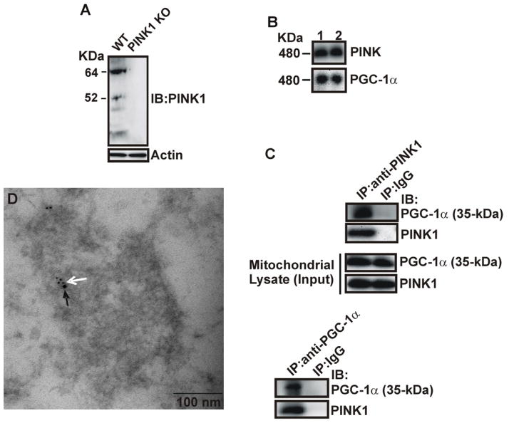
Fig. 4.
Co-localization and association of 35 kDa PGC-1α with PINK1. A) Total protein lystates from the hippocampus of PINK1 knockout and wild-type mice were analyzed by Western blotting with anti-PINK1 and actin antibodies. B) Purified mitochondria from mouse hippocampus were subjected to BN-PAGE analysis, followed by the second-dimension SDS-PAGE gels and immunoblotting with P-120 and anti-PINK1 antibodies. Lane 1&2 represent two different mice. C) Lystates from purified hippocampal mitochondria were co-immunoprecipitated with anti-PINK1, P-120, and anti-IgG antibodies, followed by immunoblotting using anti-PINK1 and P-120 antibodies. D) Immunoelectron microscopy of mitochondrial PGC-1α and PINK1 co-localization. White and black arrows indicate PGC-1α (5 nm) and PINK1 (10 nm) immunogold labeling.

