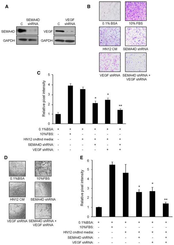
Fig. 4.
SEMA4D and VEGF are produced by HNSCC and promote endothelial cell migration and tube formation in vitro. a Immunoblot analysis for SEMA4D (upper panel, left) and VEGF (upper right) in lysates from HN12 cells infected with empty vector control lentivirus (C) or virus coding for SEMA4D shRNA or VEGF shRNA, where indicated. GAPDH was used as a loading control (lower panels). b Boyden chamber migration assay in HUVEC toward 0.1 % BSA (negative control), 10 % FBS (positive control) or serum-free media conditioned by HN12 cells, control-infected or infected with lenti-virus coding for SEMA4D shRNA, VEGF shRNA, or both. c Results of the migration assay from (b), quantified as the pixel intensity of scanned migration assay membranes relative to the negative control. Error bars represent the standard deviation from the averages from three wells (*p <0.05; **p <0.01). d HUVEC were plated on reconstituted basement membrane material in media containing 0.1 % BSA (negative control), 10 % FBS (positive control) or serum-free media conditioned by HN12 cells, control-infected or infected with lentivirus coding for SEMA4D shRNA, VEGF shRNA, or both, and examined for formation of capillary tubes. Representative photographs are shown. e Quantification of the results of the tubulogenesis assay, measuring, and summing the length of all tubular structures observed in 10 random fields. Error bars represent the standard deviation from three independent experiments (*p <0.05; **p <0.01)

