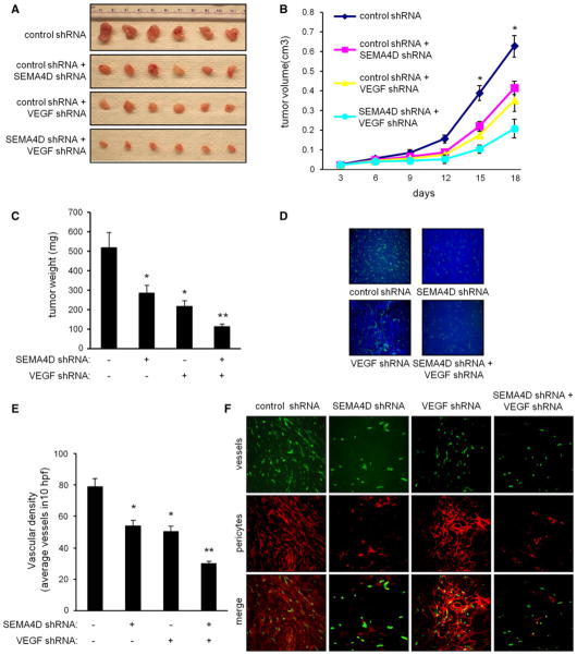
Fig. 5.
Production of SEMA4D by HNSCC cooperates with VEGF to promote tumor-induced angiogenesis. a HN12 cells infected with lentiviruses coding for control shRNA, SEMA4D shRNA, VEGF shRNA, or both were injected subcutaneously into nude mice along with a bolus of basement membrane extract. Representative tumors are shown at the time of kill. b The results of tumor volume measurement in cm3 are shown (n = 10; *p <0.05). c The results of tumor weights in mg are shown (n = 10; *, p <0.05; **p <0.01). d Immunofluorescence for CD31 as a measure of vascular density from tumors derived from HN12 cells infected with a control lentivirus (control shRNA), lentivirus-expressing SEMA4D shRNA or VEGF shRNA, or both. Nuclei are stained with DAPI. e Results of measurement of vascular content from these tumors, determined by the average number of vessels in 10 high-power fields (hpf) of CD31-stained sections, are quantified in the bar graph (*p <0.05; **p <0.01). f Immunofluorescence for vessels (green, top row) and pericytes (red, middle row) in the same tumor populations, looking for association of pericytes with endothelial cells (merge, bottom row) as an indicator of vessel integrity and maturity. Tumors from cells-expressing SEMA4D shRNA exhibit reduced numbers of vessels that fail to associate with pericytes. Tumors comprised of HN12 cells infected with VEGF shRNA-expressing lentivirus also fail to induce robust blood vessel growth, but those vessels are closely associated with a pericyte sheath

