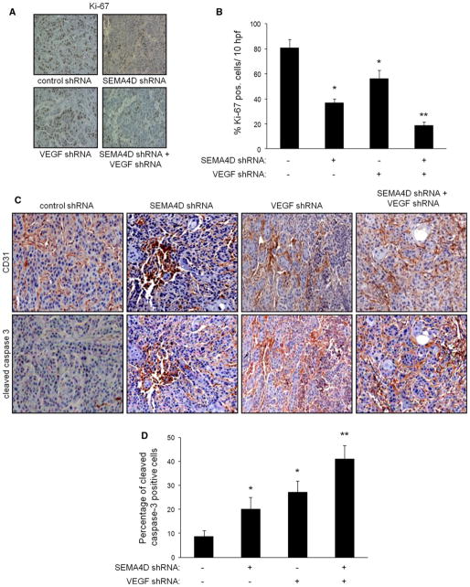
Fig. 6.
Production of SEMA4D and VEGF by HNSCC contributes to tumor cell proliferation and endothelial cell survival in tumor vasculature. a Immunohistochemistry for Ki-67, to measure proliferation of cells from tumors comprised of HN12 cells infected with control lentivirus (control shRNA), lentivirus-expressing SEMA4D shRNA, VEGF shRNA, or both. b Results of Ki-67 staining, expressed as percentage of positive cells observed from 10 high power fields. Error bars represent the standard deviation of the averages from three independent experiments (*p <0.05; **p <0.01). c The presence of cleaved caspase 3 (bottom row) was evaluated in tumors comprised of HN12 cells-expressing control shRNA, SEMA4D shRNA, VEGF shRNA or both, correlated with tumor sections stained for endothelial cells (CD31, top row). d Results of vascular apoptosis, expressed as percentage of vessels exhibiting cleaved caspase 3 observed from 10 high power fields, is shown in the bar graph (lower panel; p <0.05; **p <0.01)

