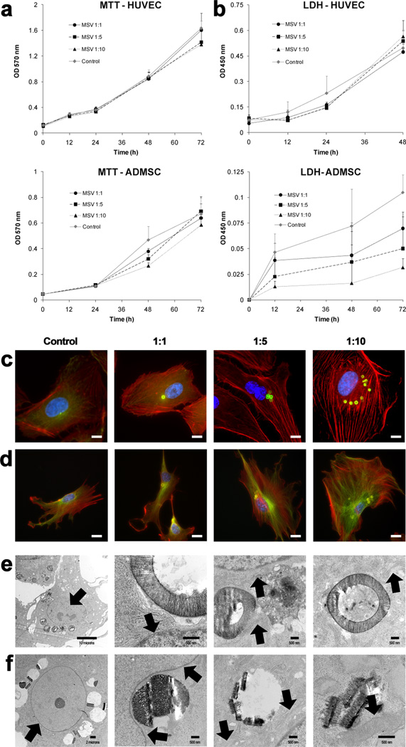
Figure 1.
Cellular proliferation, toxicity, and architecture upon internalization of MSV. A) MTT assay was used to determine the effect MSV internalization on proliferation of HUVEC (top) and ADMSC (bottom). B) LDH assay was used to assess cellular toxicity and membrane damage that could have developed within HUVEC (top) and ADMSC (bottom) after the internalization of MSV. C, D) Cytoskeleton staining of HUVEC (C) and ADMSC (D) at increasing doses (1:1, 1:5, 1:10) of MSV demonstrated conservation of cellular structure. In C & D: microfilaments (f-actin) are in red, microtubules (α-tubulin) in green, nuclei are in blue, and MSV are in green (C) or yellow (D), scale bar = 10 µm. E, F) TEM images of HUVEC (E) and ADMSC (F), 24 hours after internalization of MSV. Starting from the left, images were selected to demonstrate the effect of MSV internalization on the nucleus/nucleolus, nuclear envelope, rough endoplasmic reticulum, and mitochondria, indicated by black arrows. Scale bars are 500 nm, except for left most images where it is 10 µm (E) and 2 µm (F).

