
Fig. 2. Duodenitis in Rag1−/− recipients of CD4+CD45RBlowCD25− T cells.
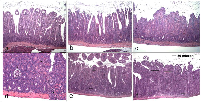
Duodenal samples were stained with H/E. a) Mild duodenitis (section taken from mouse in the ovalbumin/gluten group). Mild infiltration of villus lamina propria with mononuclear cells and presence of cryptitis, but normal crypt/villus ratio (magn. 100x). b) Moderate duodenitis (from gliadin/gfd group). Increased infiltration of the villus and basal lamina propria with mononuclear cells, presence of cryptitis (but no crypt abscesses), increase of crypt/villus ratio. c) Severe duodenitis (from gliadin/gluten group). Loss of villus structure due to severe infiltration of the lamina propria with mononuclear cells, pronounced villus shortening and elongation of crypts, cryptitis (crypt abscesses present at other locations of the same section). d) Severe duodenitis (from gliadin/gluten group; magn. 200x) with cryptitis, crypt abscess (↓) and multinucleated giant cell in lamina propria (→; see insert magn. 400x). e, f) Scoring of villus mononuclear cell infiltration (magn. 100 x), relating villus lamina propria diameters to crypt diameters. Examples for e) villus diameter 1–2x crypt diameter (score 2) and f) >2x crypt diameter (score 3).