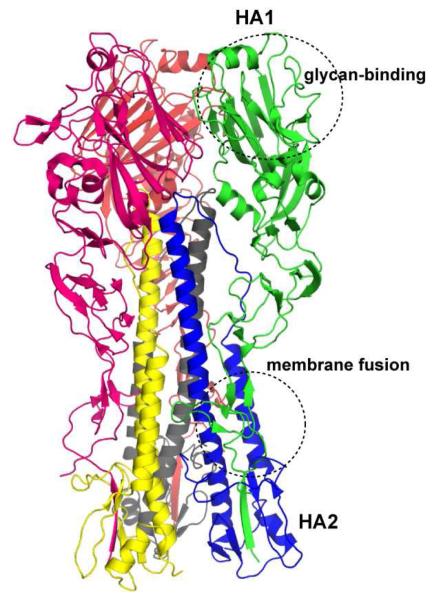
Figure 2. Structure of trimeric HA.
The cartoon rendering of a representative H1N1 HA (PDB ID: 1RU7) is shown where each chain (HA1 or HA2) in each of the monomer is colored distinctly. The region in HA1 involved in sialylated glycan receptor binding and the region between HA1 and HA2 involved in membrane fusion are also shown.

