
Figure 5.
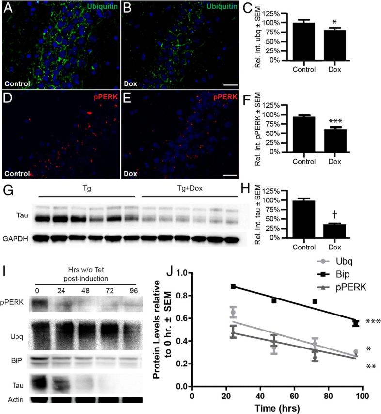
Depletion of soluble tau reverses the accumulation of ubiquitin and pPERK signal in vivo and in vitro. A–H, rTg4510 mice were fed a control or Dox diet for 35 d to suppress tau transgene expression. A, B, D, E, Representative immunofluorescent images of ubiquitin (A,B; green), pPERK (D,E; red), and DAPI (blue) in the CA2 of Tg brains (scale bar, 40 μm). C, F, Graphs showing that ubiquitin and pPERK aggregates were decreased by 20% (*p < 0.05; n = 3 control and n = 4 Dox) and 37% (***p < 0.0001; n = 5 control and n = 6 Dox), respectively. G, H, Representative immunoblot of tau from brains of Dox-fed mice and controls showing that tau levels were decreased by 64% (†p < 0.0001; n = 6 for each condition). I, Representative Western blot of pPERK, BiP, ubiquitin, tau, and actin from iHEK-Tau cells. iHEK-Tau cells were induced to express tau by adding Tet to the media. After 4 d, Tet-containing medium was removed, cells were washed, and Tet-free media was added for 4 d. Cells were harvested at different time points after removing the Tet. J, Linear regression analysis showing reductions in of pPERK, BiP, and ubiquitin levels over time after Tet removal; values are relative and normalized to actin. Significance was derived from four experiments (*p < 0.05, **p < 0.01, ***p < 0.001).