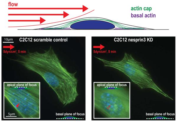
Fig. 4. LINC-mediated formation of the actin cap in cellular mechanotransduction.
Schematic of an adherent cell exposed to fluid flow shear force. Left, micrograph shows a serum-starved control C2C12 mouse myoblast cell (which contains no organized actin structures before stimulation) that forms an organized actin cap (inset) in response to exposure of a shear stress of 5 dyn/cm2 for 5 min. The right micrograph shows a serum-starved C2C12 cell that has been shRNA-depleted of LINC complex protein nesprin3. This cell is unable to form an organized actin cap after exposure to the same fluid shear stress.

