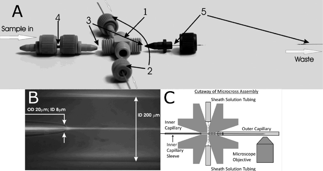
Figure 1.
(A) Mixer design. The components include: (1) MicroCross that holds 360 µm outer diameter tubing and has a 150-µm through-hole (2) 1/32” PEEK tubing that has a 250 µm inner diameter and carries sheath flow from HPLC pumps (3) Fused silica capillary for sample flow: outer diameter = 90µm; inner diameter = 20µm (4) MicroTight tubing sleeve (5) Fused silica capillary for sheath flow: outer diameter = 350 µm; inner diameter = 200 µm. All parts are connected via fitting nuts and MicroFerrules with 0.025” diameters. (B) Overlay of bright field and fluorescence images of capillary flow system. The sample flows from left to right through the inner capillary, and the sheath solution flows from left to right through the outer capillary, with dimensions as indicated. The tapered capillary tip and 3D focusing by the sheath solution create a narrow sample jet that is 2–5 µm in diameter. A laminar flow of the sample is established and rapid mixing occurs via diffusion. The sample appears as a bright stream due to fluorescence of Eu beads. The sample flow rate is 0.025 µL/min, and the sheath flow rate is 100 µL/min. (C) Cutaway of MicroCross assembly showing how solutions are kept separate (not to scale). Ferrules (not shown) hold each tube or capillary in place. The sheath solution can only flow out through the outer capillary, whereas the inner capillary outlet is outside of the cross and therefore the mixing region is outside of the cross, in the vicinity of the microscope objective.

