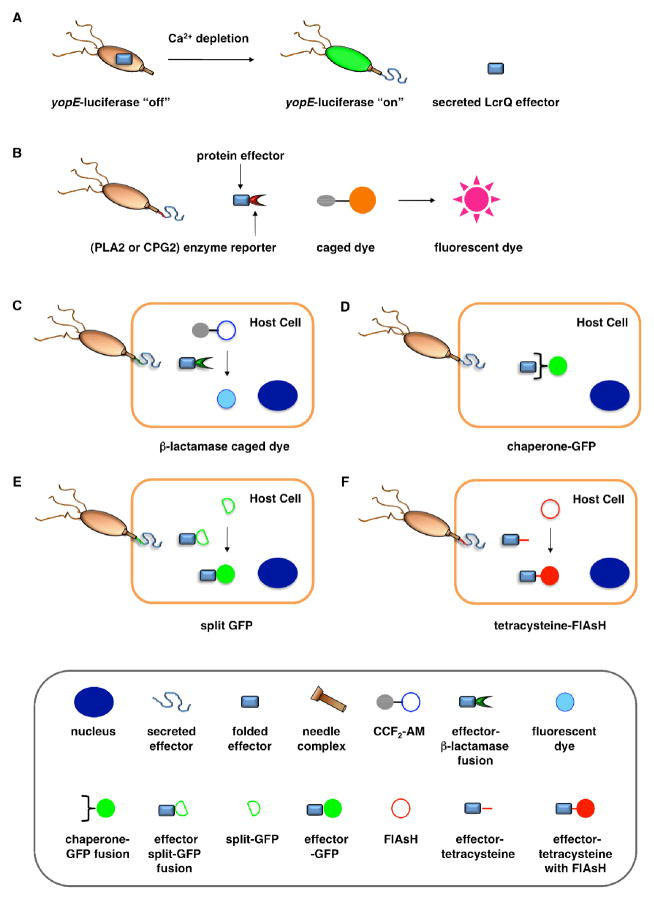
Figure 2.
Assays for bacterial effector secretion and HTS. A. Whole cell HTS using a Yersinia pseudotuberculosis (yopE:luxAB) strain21 that utilizes a transcriptional readout linked to secretion. B. A whole-cell HTS was performed by engineering a Salmonella strain where the secreted effector SipA was fused to a portion of a Yersinia protein, YplA, which contained phospholipase A2 (PLA2) activity41. Carboxypeptidase G2 (CPG2)-based reporter system for fluorescence and visible detection of type III protein secretion from Salmonella38. C. The bacterial effectors are fused with β-lactamase (βla) that cleaves a βla-sensitive FRET probe, CCF2-AM, in the host cells. D. GFP-labeled chaperones were used as probes to visualize translocation of bacterial effectors by imaging of effector accumulation in the cytosol of the host cells. E. Upon T3SS effector translocation, the association of the two fragments, small 13-amino-acid 11th strand of the GFP β-barrel and the complementary fragment of the first 10 GFP strands, leads to GFP fluorescent tagging of the effector population in the host cells. F. The fluorescein-based biarsenical dye FlAsH in the host cells allow the labeling of effectors with a genetically encoded sequence containing the tetracysteine repeat motif as the tag. Legends for C-F are indicated in the bottom grey box.

