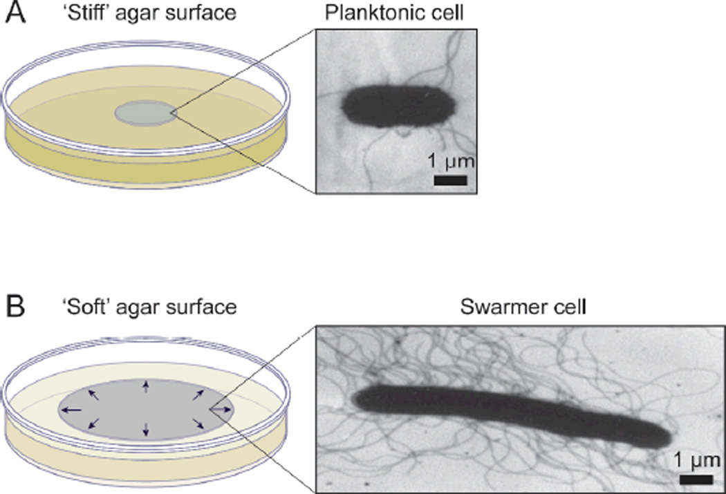
Figure 4.
A) A schematic diagram depicting a colony of bacteria grown on the surface of a ‘stiff’ agar gel (e.g. 1.5%, w/v) and a characteristic planktonic cell isolated from the colony. The TEM image shows a planktonic cell of S. liquefaciens MG1.83 B) A swarming colony of bacteria grown on the surface of a ‘soft’ agar gel (e.g. 0.45%, w/v) and a TEM image of a swarmer cell of S. liquefaciens MG1 isolated from the outer edge of a swarm colony.83 The arrows depict the radial outward expansion of the colony on the surface. TEM images in A and B are reprinted with permission from the American Society for Microbiology, Copyright 1999.

