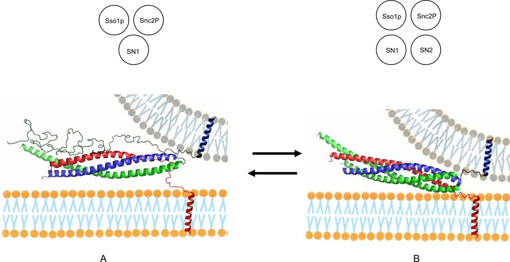
Figure 3. Models for the Partially Assembled SNARE Complex and the Fully Assembled Four–Helix Bundle.
(A) Partially assembled complex with the C-terminal region of Sso1pHT and the entire length of SN2 of Sec9c unstructured. (Inset) a diagram for a three-stranded coiled coil composed of Sso1p, SN1 and Snc2p.
(B) Fully assembled four-helix bundle. The EPR analysis show that A and B are nearly equally populated. The SNARE proteins are color-coded: red, Sso1p; green, Sec9c; blue, Snc2p. The apposing membrane in gray is drawn arbitrarily to help speculate the trans SNARE complexes. (Inset) a diagram for a four-stranded coiled coil composed of Sso1p, SN1, SN2, and Snc2p. Both partially and fully assembled complexes were constructed based on the crystal structure (Sutton et al., 1998) with PyMOL.

