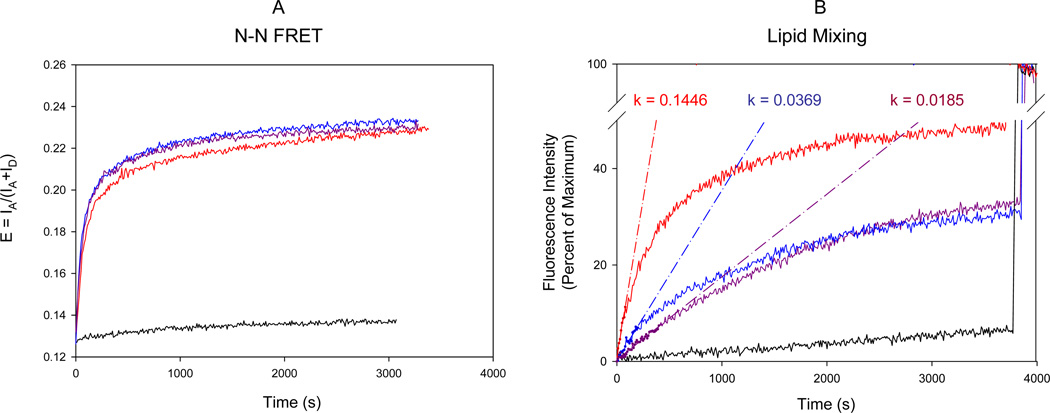
Figure 4. FRET Assays of SNARE Assembly and Membrane Fusion.
(A) FRET assay of trans SNARE assembly and vesicle docking. The fluorescence change was monitored in two channels with the excitation wavelength of 545 nm and the emission wavelengths of 570 and 668 nm, respectively. FRET efficiency E was defined as E = IA/(IA+ID). Little fluorescence change was observed without Sec9c (black line). The reaction was initiated by adding wild type Sec9c (red line) or its proline mutants (L626C, blue line; L647C, purple line) to the mixture of t-SNARE vesicles containing Cy3-labeled Sso1pHT and the v-SNARE vesicles carrying Cy5-labeled Snc2p. Labels were attached near the N-terminal tips.
(B) Fluorescence lipid mixing assay of SNARE-mediated membrane fusion. Fluorescence changes for lipid mixing were normalized with respect to the maximum fluorescence intensity (MFI) obtained by adding 0.1% reduced triton x-100. The black line represents the negative control in the absence of Sec9c. The red, blue and purple lines represent the fluorescence changes after adding wild-type Sec9c, L626P and L647P, respectively. The k values represent the initial rate of the fusion kinetics in units of percent per sec.

