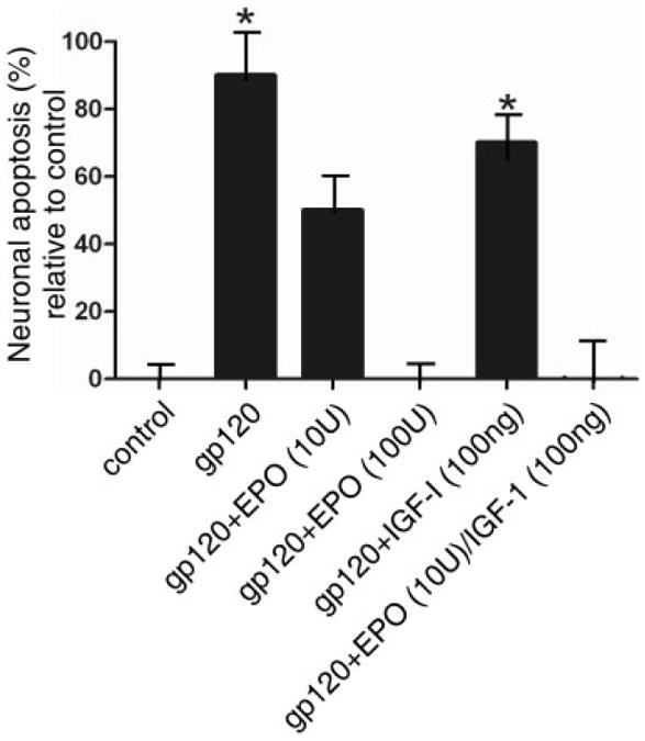
FIGURE 2.

Neuroprotection of rat cerebrocortical neurons from gp120-induced apoptosis, showing protection by erythropoietin (EPO), insulin-like growth factor-I (IGF-I), or a combination of both. Mixed neuronal/glial cerebrocortical cell cultures were exposed for 24 hours to gp120 (200pM) in the presence or absence of the indicated concentrations of EPO and IGF-I. Each experiment was performed in triplicate and repeated at least 3 ×. Approximately 1,000 neurons were scored for each value. The y (ordinate) axis represents the percentage of neurons undergoing apoptosis above control levels. Approximately 20% of the neurons not exposed to gp120 died by apoptosis in the control cultures, as expected among developing neurons. *p < 0.01 by ANOVA with post hoc Scheffé test.
