
FIGURE 6.
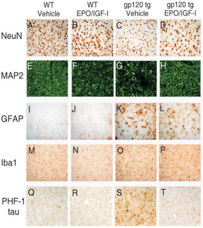
Neuropathological characterization of the protective effect of erythropoietin (EPO)+insulin-like growth factor-I (IGF-I) against gp120 toxicity in the mouse cortex. Brain sections of 10-month-old wild-type (WT) and gp120-transgenic mice treated chronically with EPO+IGF-I versus control were analyzed for degree of cortical neuropathology. Serial 40μm brain sections were immunostained for NeuN (A–D), microtubule-associated protein-2 (MAP-2) (E–H), glial fibrillary acidic protein (GFAP) (I–L), Iba1 (M-P), or phospho-tau (PHF-1) (Q-T). Note the loss of NeuN and MAP-2 staining in gp120-transgenic mice (C and G), but NeuN and MAP-2 staining approached WT levels after chronic treatment with EPO+IGF-I (D and H). GFAP and Iba1 staining in gp120-transgenic mice did not change after EPO+IGF-I treatment. Human immunodeficiency virus/gp120-transgenic mice manifested increased PHF-1 tau staining (S) compared to WT (Q) or WT treated with EPO+IGF-I (R). PHF-1 tau was dramatically reduced in gp120-transgenic mice after chronic treatment of EPO+IGF-I (T).