
FIGURE 7.
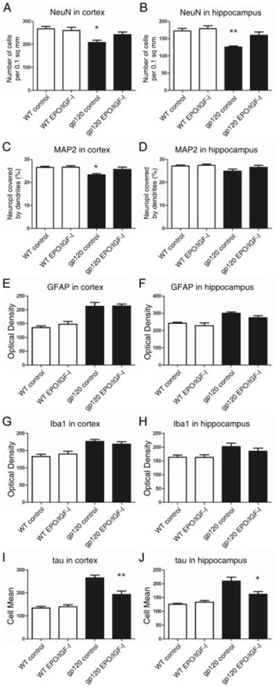
Computer-aided image analysis of neurodegeneration in the cortex and hippocampus of wild-type (WT) and human immunodeficiency virus/gp120-transgenic mice treated with erythropoietin (EPO)+insulin-like growth factor-I (IGF-I). Laser scanning confocal images of immunolabeled neocortical (A, C, E, G, and I) and hippocampal sections (B, D, F, H, and J; 5–7 mice per group) were analyzed quantitatively using NIH Image. Representative laser scanning confocal images revealed significant increases in NeuN (A and B) and microtubule-associated protein-2 (MAP-2) (C), but a decrease in phospho-tau (I and J) in gp120-transgenic mouse brains treated with EPO+IGF-I compared to vehicle. Values represent mean + standard error of the mean. *p < 0.05, **p < 0.005, by 1-tailed Student t-test comparing vehicle versus EPO+IGF-I–treated gp120-transgenic mice. GFAP = glial fibrillary acidic protein.