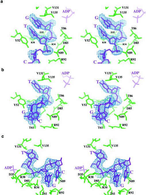
Figure 1.
Composite omit maps of the DNA binding region and kinase active site. (a) Observed density for the 5′-GTCAC-3′ substrate. The second base (thymine) is stacked against the 5′ guanine and forms a polar aromatic interaction with Y52. The edges of both bases are solvent-exposed, and the opposite face of the guanine base is flanked by non-polar residues in the base of the active site. (b and c) The density for the 5′-TGCAC-3′ substrate [(b) is in the same orientation as (a); (c) is rotated 90° relative to (b) to better visualize the stacking interaction between the second and third bases]. The 5′ thymine base and its ribose sugar are located in a similar position as the 5′ guanine on the previous panel. The second base (guanine) has swung out of the active site cleft and is stacked against the third base (cytosine). The structural roles of several side chains in DNA binding are different between the two complexes.

