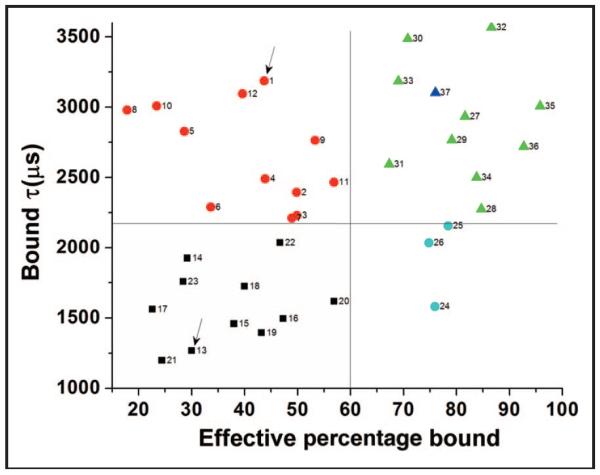
Fig. 4. Identification and classification of vWD with 2-component FCS fits.
Type 1 vWD patients ( ) have a distribution of mean diffusion times that overlaps that of samples with vWF within reference intervals (
 ), but with a significantly reduced effective bound fraction, reflecting reduced amounts of a relatively normal distribution of multimers in type 1 vWD. Type 2 vWD patients have an effective bound fraction component that overlaps that of type 1 vWD patients but with a reduced diffusion time, reflecting a lack of large multimers (
 ). Three samples with high vWF:Ag but abnormal multimer analysis lacking large multimers (
 ) show a high effective bound fraction and short mean diffusion time. Two of these samples have been classified clinically as acquired vWD. The type 1 and type 2 vWD samples shown in Fig. 3 are denoted by arrows. The 4-h post-DDAVP sample described in Fig. 5 is also shown for reference (
 ).
).

