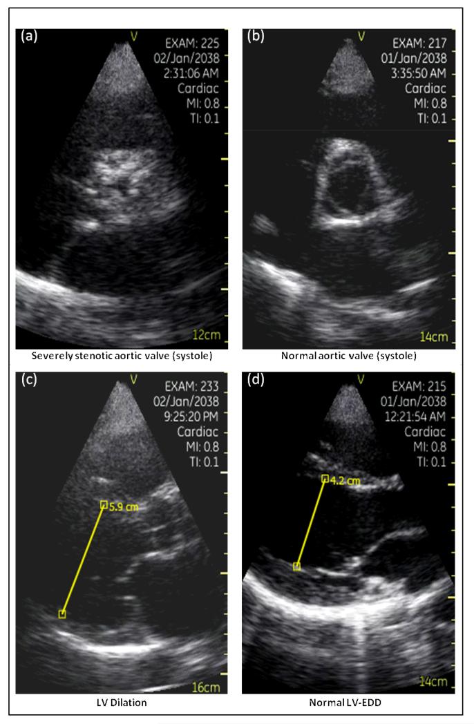
Figure 2.
Pocket-mobile echo (PME) images of (a) severely stenotic aortic valve and (b) a normal aortic valve, both images recorded from a parasternal short-axis view during ventricular systole. PME images of (c) enlarged LVEDD and (d) a normal LVEDD in the parasternal long axis view as measured by electronic calipers built into the ultrasound device’s software.

