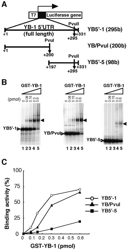
Figure 6.
Identification of the YB-1 binding region in YB-1 5′-UTR mRNA. (A) Schematic illustration of YB-1 5′-UTR deletion constructs used as the probe in a REMSA. To produce RNA of defined length, restriction enzyme (PvuI or PvuII) was used to linearize the DNA templates. (B) REMSA. The indicated amount of GST–YB-1 fusion was incubated with each 32P-labeled YB-1 5′-UTR deletion mRNA at 25°C for 15 min. An arrow indicates the YB-1/YB-1 5′-UTR RNA complex. YB-1/YB-1 5′-UTR RNA probe complexes were separated using 4% native polyacrylamide gels. (C) Kinetic analysis of GST–YB-1 binding to YB5′-1, YB/PvuI and YB5′-5 probes. The GST–YB-1 binding activity to YB-1 5′-UTR fragments identical to that shown in (B) were quantified by monitoring amounts of the binding complexes.

