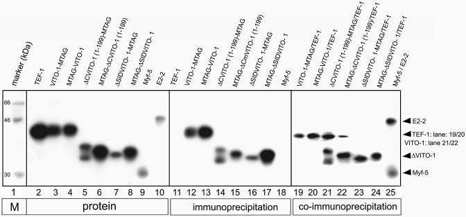
Figure 1.
Interaction of VITO-1 with TEF-1 depends on the SID domain. SDS–gel electrophoresis of in vitro translated, [35S]methionine-labelled TEF-1 (lanes 2), VITO-1-MTAG (lane 3), MTAG-VITO-1 (lane 4), ΔCVITO-1(1–199)-MTAG (lane 5), MTAG-ΔC-VITO-1(1–199) (lane 6), ΔSID-VITO-1(108–323)-MTAG (lane 7), MTAG-ΔSID-VITO-1(108–323) (lane 8), Myf-5 (lane 9) and E2-2 (lane 10). Control immunoprecipitations with an anti-myc epitope antibody (lanes 11–17) and an anti-E2-2 antibody (lane 18). Co-immunoprecipitations of unlabelled VITO-1-MTAG together with labelled TEF-1 (lane 19), unlabelled MTAG-VITO-1 together with labelled TEF-1 (lane 20) and labelled TEF-1 (lanes 21–24) together with labelled ΔCVITO-1(1–199)-MTAG (lane 21), MTAG-ΔCVITO-1(1–199) (lane 22), ΔSID-VITO-1(108–323)-MTAG (lane 23) and MTAG-ΔSID-VITO-1(108–323) (lane 24). A control co-immunprecipation of E2-2- and Myf-5 with an antibody against E2-2 is shown in lane 25. The co-immunoprecipitation clearly demonstrates that VITO-1 and TEF-1 form a complex in the absence of DNA.

