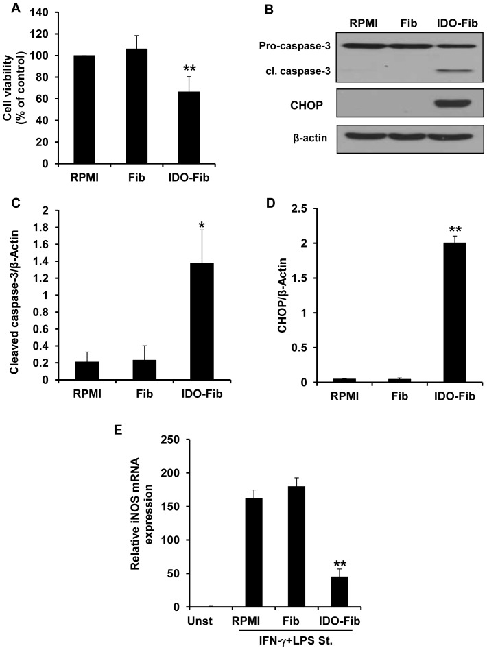
Figure 6. Effect of IDO expression by bystander fibroblasts on viability and proinflammatory activity of peritoneal macrophages.
(A) Evaluation of the effect of IDO induced tryptophan deficiency/kynurenine accumulation on primary macrophage viability. Peritoneal macrophages were either cultured in RPMI or co-cultured with control (Fib) or IDO-expressing fibroblasts (IDO-Fib). MTT assay was done after 3 days of incubation. (B) Apoptosis induction in macrophages co-cultured with IDO-expressing fibroblasts. Presence of cleaved caspase-3 (cl. caspase-3) and CHOP was analyzed in macrophage cell lysate following 3 days of co-culture. The ratio of cleaved caspase-3/β-actin (C) and CHOP/β-actin (D) expression in macrophages, respectively. (E) Inhibition of proinflammatory activity in peritoneal macrophage co-cultured with IDO-expressing fibroblasts. iNOs expression was evaluated in INF-γ+LPS stimulated macrophages preincubated in PRMI or conditioned medium collected from either control (Fib) or IDO-expressing fibroblasts (IDO-Fib). GAPDH was used as the reference gene. Data is mean±SEM of three independent experiments (*P-value<0.05, **P-value<0.01, n = 3).

