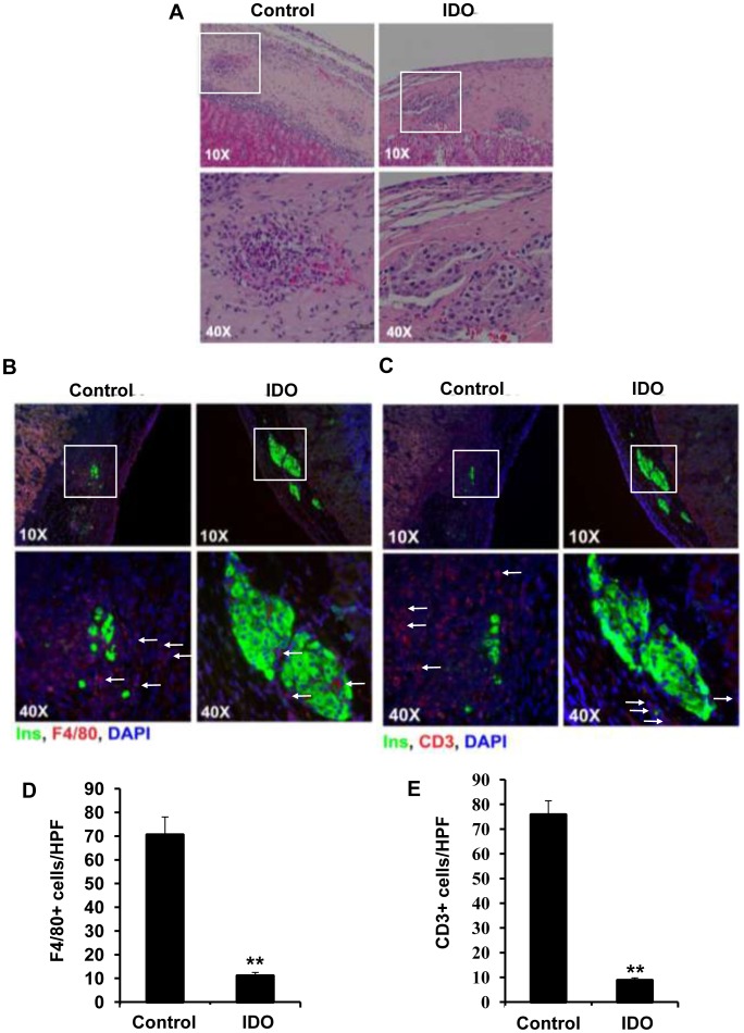
Figure 7. Infiltration of composite islet xenografts by F4/80+ and CD3+ cells.
Three dimensional grafts were constructed by embedding isolated rat islets in the collagen matrix populated with IDO-expressing or control B6 mouse fibroblasts. Graft-recipient mice were killed 10 days after transplantation of the composite xenografts. Retrieved composite islet grafts were then subjected to H&E staining (A) and double immuno-fluorescence staining for F4/80 and insulin (B) or CD3 and insulin (C). The arrows indicate infiltrated immune cells. Quantitative analysis of F4/80+ macrophages (D) and CD3+ T cells (E) infiltration into islet xenotransplants. Graphs show manual counting scores±SEM per high-power field (HPF, x400). Data derived from the examination of four different HPFs per tissue section (**P-value<0.01, n = 4).

