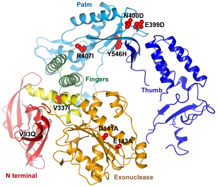
Figure 3. Structure of the Pfu-E10 polymerase.
Cartoon representation of the apo polymerase structure with domains coloured as follows: N-terminal domain: red, exonuclease domain: gold, linker region: yellow, palm domain: pale blue, fingers domain: green and thumb domain: dark blue. The side chains of the mutated residues (V93Q, D141A, E143A, V337I, E399D, N400D, R407I and Y546H) are shown with atoms represented as red spheres.

