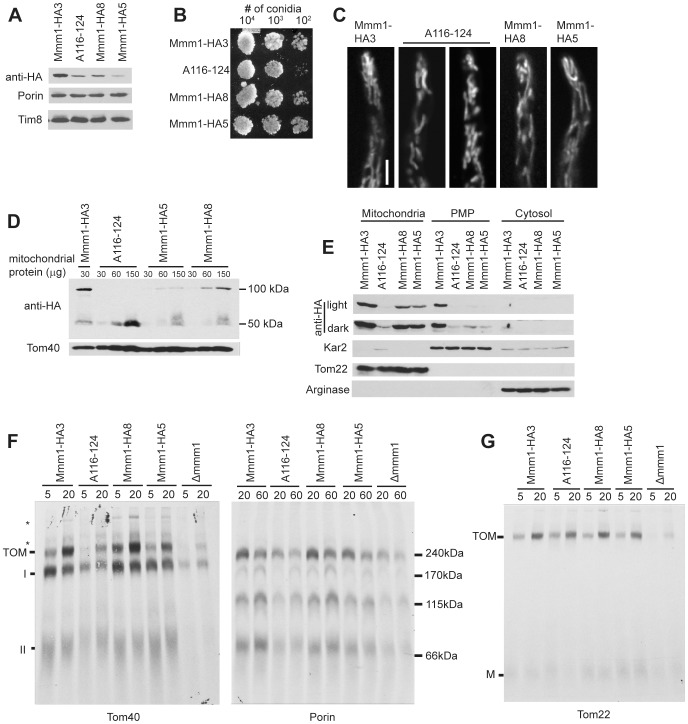
Figure 5. Characterization of N. crassa Mmm1 conserved region mutant A116-124.
A. Western blot analysis of crude mitochondria (30 ug) from the indicated strains. Samples were subjected to SDS-PAGE, transferred to nitrocellulose and analyzed by Western blotting for the indicated proteins. B. Measurement of growth as in Figure 3C. C. Confocal microscopy of mitochondria as in Figure 3D. D. Non-reducing SDS-PAGE followed by Western blot analysis as in Figure 3E, but 30, 60, and 150 µg of mitochondria isolated from the indicated strains were loaded. E. Cell fractionation as in Figure 1A. F. Assembly of radiolabeled β-barrels as in Figure 4A using crude mitochondrial preparations as indicated in the Methods. G. Assembly of radiolabeled Tom22 as in Figure 4C.

