
Figure 6. Characterization of the mmm2 knockout strain.
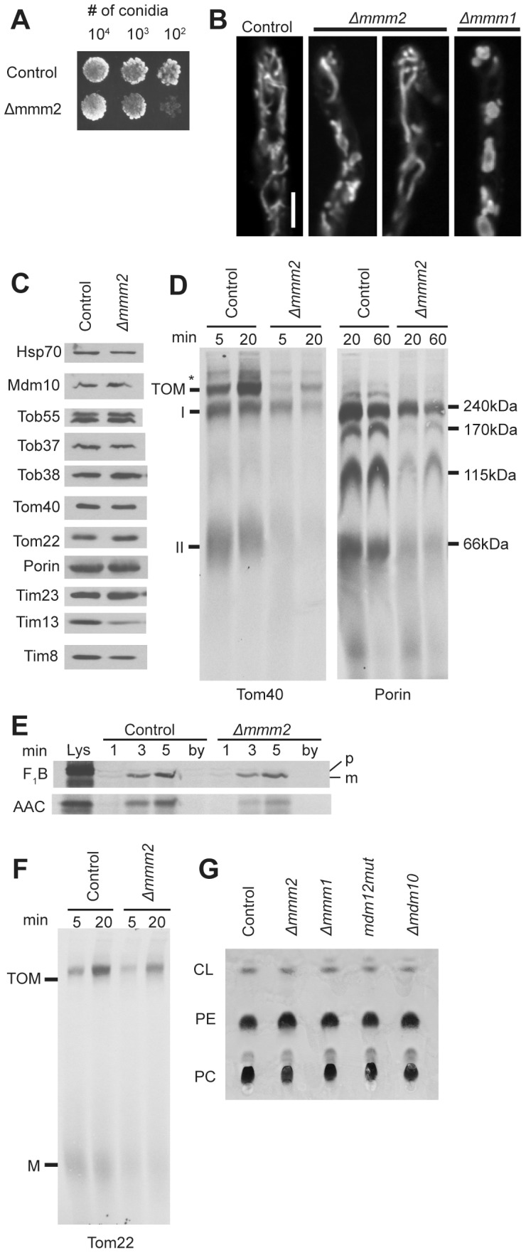
A. Measurement of growth rate as in Figure 3C. B. Visualization of mitochondria as in Figure 3D. C. Mitochondria (30 ug) isolated from the control and mutant strains were subjected to SDS-PAGE, transferred to nitrocellulose and analyzed on Western blots with antibodies to the indicated proteins (30 µg mitochondrial protein per lane). D. Assembly of radiolabeled β-barrel proteins Tom40 and porin as in Figure 4A and B, respectively. E. Radiolabeled F1β and AAC were imported into mitochondria isolated from the indicated strains. After import, the mitochondria were treated with proteinase K, reisolated, electrophoresed, transferred to nitrocellulose membranes, and examined by autoradiography. Lys, 33% of the input lysate containing radiolabeled protein used in each reaction; by (bypass import), mitochondria pretreated with trypsin to remove surface exposed receoptor proteins before 3 min of import with precursor proteins. This lane serves as a control to show no import occurs when mitochondrial surface receptors have been removed; p, preprotein; m, mature protein. F. Assembly of radiolabeled Tom22 as in Figure 4C. G. Total mitochondrial lipids were extracted from isolated crude mitochondria (300 µg protein) from the indicated strains in 1:1 chloroform : methanol. Lipids were then analyzed by TLC, stained with molybdenum blue, and photographed. PC, phosphatidylcholine; PE, phosphatidylethanolamine; CL, cardiolipin.