
Figure 2.
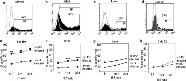
Efficiency of H-1PV infection and NK-cell-mediated killing of mock- and H-1PV-infected colon carcinoma cells. Colon carcinoma cells were buffer-treated or infected at MOI=5 RU/cell with recombinant H-1PV expressing the marker EGFP (Chi-hH1/EGFP). The proportion of cells expressing EGFP was determined by flow cytometry. Graphs a-d: gray lines: autofluorescence profiles of mock-treated cells; black columns: specific staining profiles of Chi-hH1/EGFP-infected. Graphs e-h: Colon carcinoma cells were infected with H-1PV (MOI=5RU/cell) or mock-treated, incubated for 24 h, and labeled with 51Cr for 1 h. Labeled cells were then incubated with IL-2-activated NK effector cells (E) for 4 h at the indicated E:T ratio, and cell lysis was measured (Figure 3e-h). The data shown are means with SD bars of the results corresponding to NK cells from 4 different donors, each measurement being performed in triplicate.