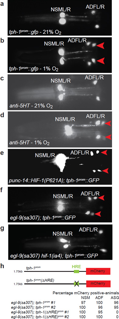
Fig. 1.
5-HT expression is altered in hypoxia through direct HIF-1 regulation of tph-1
(A–B) tph-1prom∷gfp expression in normoxia (A) and after exposure to 1% O2 for 12 hours (B). Using a reporter strain that expresses tph-1prom∷gfp at low-levels (injected at 2ng/µl) we found that tph-1prom∷gfp is also upregulated in the ADF and NSM neurons after hypoxic exposure (see Suppl. Fig. 1). (C–D) Anti-5-HT immunofluorescence of a normoxic animal (C) and an animal after exposure to 1% O2 for 12 hours (D). (E–G) tph-1prom∷gfp is ectopically expressed in ASGL/R under conditions that stabilize hif-1, achieved through either mutating the residue in HIF-1 required for its degradation (E) or in egl-9 mutants (F); the latter effect genetically depends on hif-1 (G). Red arrowheads point to the ASG chemosensory neurons. Ventral views, anterior to the left. (H) HIF-1-induced tph-1 reporter gene expression (induction achieved through preventing the degradation of HIF-1 in egl-9 mutants) is abolished upon deletion the canonical HIF-1 binding site (“HRE”'; 6 bp deletion). # = independent transgenic lines (n = 87–122).

