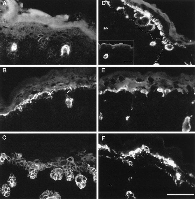
Figure 6.
Immunofluorescence analysis of wound-healing keratin expression in skin of wild-type (A–C) and K5−/− mice (D–F). In wild-type mice, K6 expression was restricted to hair follicles and single suprabasal cells (A), whereas it was strongly increased in blisters of K5−/− mice (D). In the blister, hair follicles torn out of the dermis were noted. In unaffected areas, K6 induction was comparable to the wild type Bar, inset, 60 μm. The dotted line denotes the position of the blister base. The expression of K16 (E) was not changed remarkably when compared with the wild type (B). Note that the K17 expression in the wild-type skin (C) was almost comparable to K14 staining. In K5−/− mice, K17 (F) did not show a significant increase in expression. We noted, however, the same punctuate staining as observed before for K14 and K15. Bar, 50 μm.

