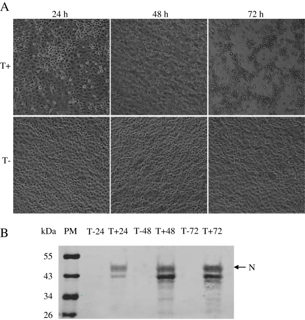
Figure 1.
Cytopathic effect caused by TGEV infection and identification of the TGEV infected and non-infected ST cells. The cytophatic effect induced by the TGEV virus was analyzed by optical microscopy, at 24 h, 48 h and 72 h p.i. Images were taken with a 40× objective (A). The N protein in TGEV-infected and mock-infected ST cells were checked using the mAb to N protein of the TGEV by the method of Western blot (B). T + and T- represent the TGEV infected and uninfected ST cells, respectively.

