Figure 1.

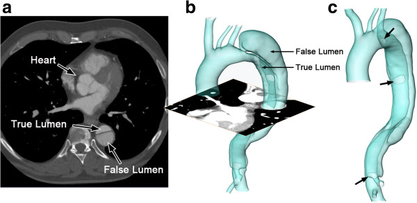
The reconstructed surface of the aortic dissection. (a) is one axial slice of the CT scan; (b) is the reconstructed surface of the aortic dissection; (c) shows the positions of the entries along the flap (indicated by arrows).

The reconstructed surface of the aortic dissection. (a) is one axial slice of the CT scan; (b) is the reconstructed surface of the aortic dissection; (c) shows the positions of the entries along the flap (indicated by arrows).