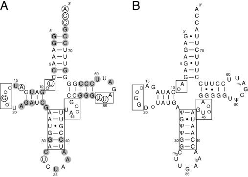
Figure 1.
Cloverleaf structure of (A) M.barkeri tRNAPyl and (B) bovine mitochondrial tRNASer(UGA). Numbering is according to Sprinzl et al. (18). Missing residues compared with canonical tRNAs are indicated by empty dots and non-canonical primary features are boxed. Consensus nucleotides in M.barkeri lysine-accepting tRNAs are in grey, whereas those conserved among all tRNAs are circled.

