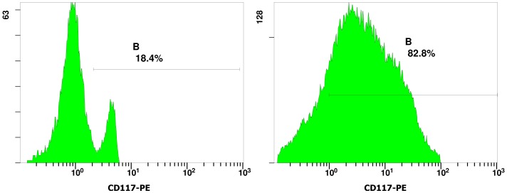
Figure 2. Immunophenotyping of CD117+ yolk sac cells.
Representative flow cytometric data from more than three independent analyses were shown. The cells were fluorescently stained with CD117-PE. To show the CD117+ cell purity, CD117+ yolk sac cells before separation were shown on the left panel; CD117+ yolk sac cells after separation by using the anti-murine CD117 antibodies conjugated to mini-magnetic beads were shown on the right panel.

