
Figure 10. DSB repair induced by HQ in BM-HSC.
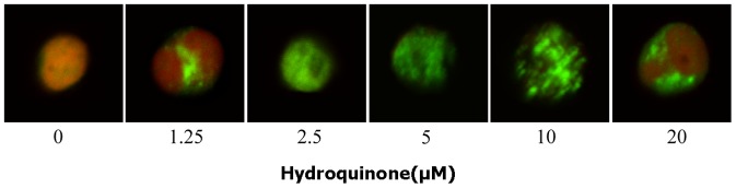
Representative microscopic fluorescence images of γ-H2AX foci accumulation after HQ treatment for 6 h. Nuclei were stained using PI and Al-exa488-labeled secondary antibodies, and were examined by confocal microscopy (oil, 200×). The experiments were repeated for three more times. Green fluorescence indicates formation of γ-H2AX foci.