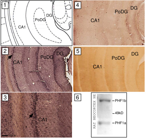
Figure 12.
Immunodetection of PHF1 proteins in nuclear extracts of primary rat neocortical cultures and slices of adult rat hippocampus. Western analysis of PHF1a and PHF1b expression using nuclear extracts of rat neocortical neurons and a primary antibody raised against a PHF1 peptide present in both PHF1a and PHF1b (panel 6). Relative size of PHF1 proteins is as indicated using relationship of migration pattern of putative PHF1a and b to position of marker proteins. Adult rat brains were sectioned coronally at Bregma −6.3mm (as depicted in panel 1) and treated with a primary antibody to PHF1 as described above. Positive immunostaining is indicated by brown-black precipitates (panel 2 and 3). Regions of CA1 (field CA1 of hippocampus), DG (dentate gyrus) and PoDG (polymorph layer dentate gyrus) are indicated as references. Panel 2 displays high PHF1 immunoreactivity in the CA1 region. Arrow in panel 2 indicates area of CA1 region that was magnified (100×) in the display of panel 3. A dark scale bar at the bottom of panel 3 shows the virtual distance between two points. Hippocampal slices processed in parallel to the experimental were treated with the PHF1 antibody blocking peptide and secondary antibody (shown in panel 4). A representative hippocampal slice processed in parallel treated only with the secondary antibody is displayed in panel 5 as an additional control.

