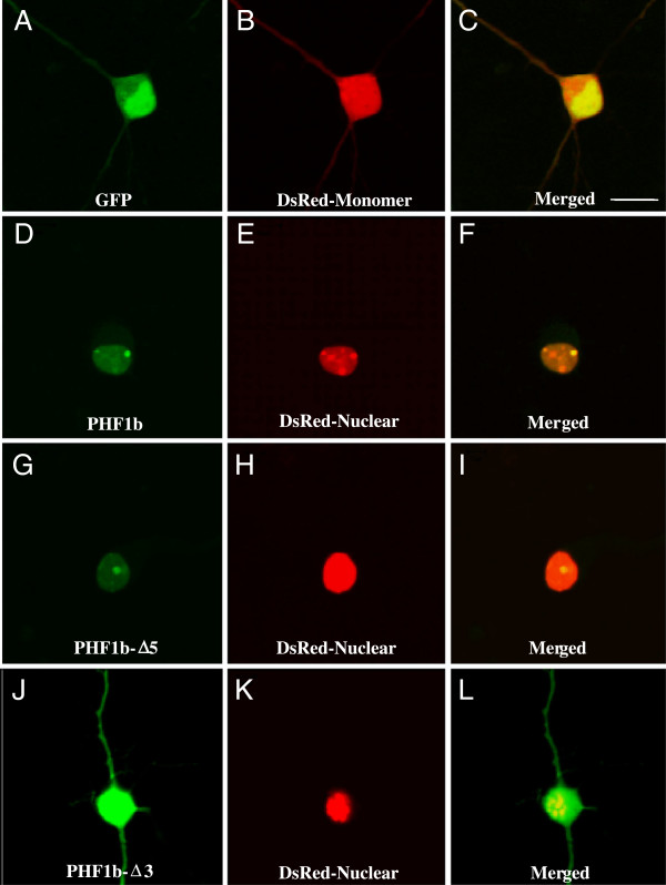
Figure 5.
PHF1b protein nuclear localization in rat neocortical neurons. Primary rat neocortical neurons isolated from E18 brain and maintained one week in vitro were transfected with GFP-PHF1b fusion plasmids (CMV-GFP-PHF1b, CMV-GFP-PHF1b-Δ5, CMV-GFP-PHF1b-Δ3, see Figure 3) and examined 48 hours after transfection by confocal microscopy for nuclear localization relative to DsRed-Nuclear marker (CMV-DsRed-Nuclear), a red fluorescent protein that localizes to the nucleus. Control transfection of CMV-GFP (A, C) and CMV-DsRed-Monomer (B, C) construct expression is throughout cortical cells and expression is not restricted to the nucleus (C). Both GFP-PHF1b (D, F) and GFP-PHF1b-Δ5 (G, I) fusion construct expression coincides with DsRed-Nuclear (E, F and H, I) indicating that both the PHF1b and the PHF1b-Δ5 protein contain a nuclear localization signal. In contrast, the GFP-PHF1b-Δ3 (J, L) fusion construct expression is not restricted to the nucleus (DsRed-Nuclear, K, L) suggesting that the nuclear localization signal of PHF1b is not localized at the N-terminus of the PHF1b protein. Scale bar is 10 μm.

