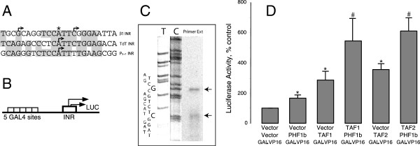
Figure 9.

Study of the β1-INR in COS cells. A) Sequence similarities between β1-INR, TdT INR and the adeno-associated virus (AAV) P5+1 INR. Shadowed boxes highlight sequence identity. Arrows indicate transcription start sites. Star symbol indicates the major start site of β1-INR in neocortical neurons (7). B) Depiction of a TATA-less synthetic promoter/reporter construct carrying a single β1-INR with a GAL4 UAS (p5XG-β1-INR-Luc). Promoter activity of the construct is regulated by co-expression of an upstream activator (GAL4-VP16). Arrows show the direction of transcription. C) Primer extension analysis of RNA from COS-7 cells transfected with the p5XG-β1-INR-Luc construct. Primer extension products are separated on a sequencing gel. Sequencing reactions (dC and dT) were run alongside of primer extension products to determine the exact start sites for initiation. Top strand DNA sequence of β1-INR is also shown next to sequencing lanes. Arrows indicate the major initiation sites in COS-7 cells. D) Co-activation of PHF1b transcriptional activity by TAF1 and 2. COS-7 cells were co-transfected with p5XG-β1-INR-Luc, GAL4-VP16 and combinations of PHF1b, TAF1 and TAF2. 48 hours after transfection, cells were harvested and assayed for luciferase activity. Results shown are mean values ± SEM and normalized to protein content within each dish as well as to vector control (Vector+Vector+GAL-VP16 defined as 100%). “*” indicates significantly different from vector control (p < 0.05) as determined by 95% confidence interval. “#” indicates significantly different from PHF1b (Vector+PHF1b+GAL-VP16) (p < 0.05) as determined by 95% confidence interval.
