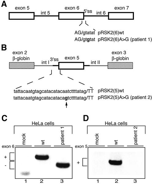
Figure 2.
Effect of the IVS6 +3 A→G and the IVS4 –11 A→G mutations in in vivo splicing assays. (A and B) Schematic representation of minigenes for RSK2 exons 6 and 5, respectively. Annotations are as in Figure 1. In (B), the genomic region of exon 5 was inserted between β-globin exons 2 and 3 represented by striped boxes. Chimeric introns upstream and downstream of exon 5, arbitrarily designated introns I and II, are 384 and 653 nt long, respectively. (C and D) RT–PCR analysis on RNA from HeLa cells transfected with exon 6 and exon 5 minigenes. In mock assays, cells were transfected with an empty vector. In (C), pRSK2(6)A→G recapitulated the splicing pattern of endogenous similarly mutated RSK2, producing complete skipping of exon 6 (lane 3). In (D), no splicing product was detectable when pRSK2(5)A→G was transfected into HeLa cells (lane 3).

