Figure 4.
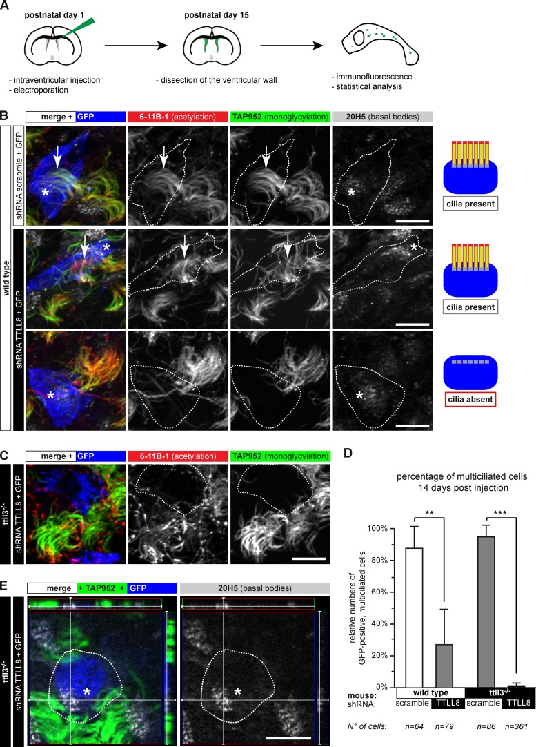
Long-term ablation of glycylases leads to nonciliated ependymal cells. (A) Flow scheme of the experimental paradigm used for shRNA-mediated depletion of TTLL8 in vivo. (B) Ependymal layer of wild-type mice. GFP-positive cells (blue or contours) express shRNA. Motile cilia were labeled for acetylation (6-11B-1), monoglycylation (TAP952), and basal bodies (20H5). Expression of TTLL8-shRNA partially led to loss of motile cilia; cells still contain multiple basal bodies. Quantification in D. (C) Expression of TTLL8-shRNA in the ependymal epithelium of ttll3−/− mice. Transfected cells (blue or contours) have no cilia. (D) Quantification of multiple motile cilia on shRNA-expressing cells 14 d after electroporation (B, C, and E). Three independent experiments per condition were performed (total number of counted cells are given below). Mean values with SEM and statistics (Welch t test) are represented (**, P < 0.01; ***, P < 0.001). (E) 3D images of TTLL8-depleted cells in the ependymal layer show fully developed and correctly arranged multiple basal bodies in cells without motile cilia. Panels on the top and the right represent the Z-stack of the image. (B, C, and E) Arrows, cilia; asterisks, basal bodies in GFP-positive cells. Bars, 10 µm.

