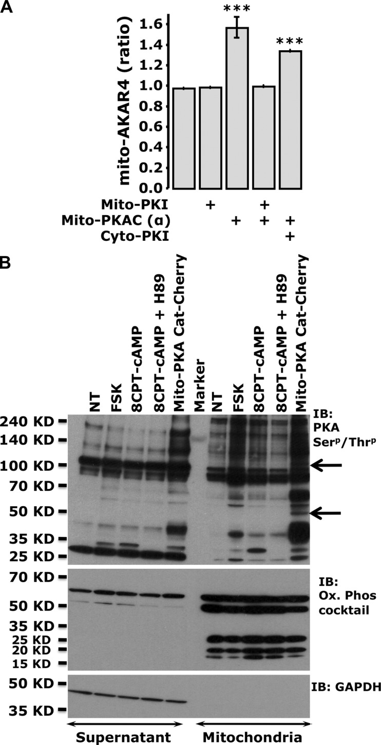
Figure 3.

Overexpression of PKA in the matrix is detected by AKAR sensors, and induces specific phosphorylation patterns. (A) Summary of the starting mito-AKAR4 ratios in the presence of mito-PKACat-mCherry, mito-PKI-mCherry, and cyto-PKI-mCherry (***, P < 0.0002 with respect to control). Error bars indicate mean ± SD. (B) Phosphorylation status of cytosolic and mitochondria-enriched fractions of HEK cells treated with cAMP-generating agonists or cell-permeant cAMP analogues, or transfected with mito-PKACat-mCherry. A phospho-(Ser/Thr) PKA substrate antibody unveiled mito-PKACat-mCherry–dependent mitochondria-specific phosphorylation bands (arrows). The purity of mitochondria was tested using an antibody cocktail against the human OXPHOS subunits, whereas GAPDH was used to detect any cytosolic contamination (typical of n = 3 independent experiments).
