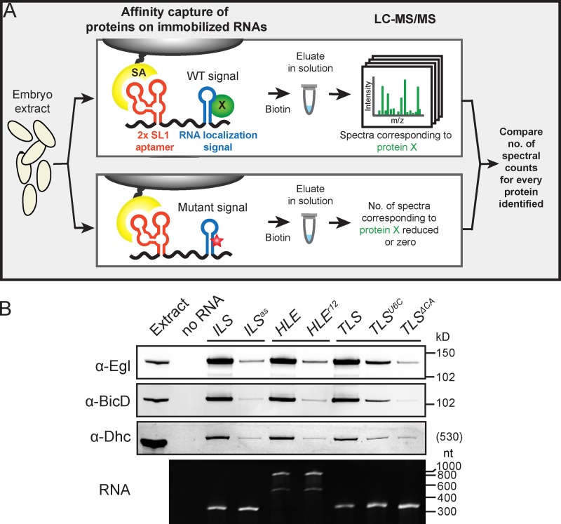
Figure 1.
A biochemical screen for novel components of mRNA transport complexes. (A) Cartoon depicting methodology used to identify novel components of mRNA transport complexes. For simplicity only one of the two streptavidin-binding aptamers in the in vitro transcribed RNA is shown. SA, streptavidin; red star, mutation in localization signal; green circle, protein enriched on WT signal. (B) Top panels, immunoblots showing that Egl, BicD, and Dhc are strongly enriched on the ILS, HLE, and TLS signals compared with mutated nonlocalizing equivalents (ILSas, HLEr12, TLSU6C, and TLSΔCA). Bottom panel, ethidium bromide–stained TBE-urea gel showing equivalent amounts of WT and mutant RNAs after coupling to beads, washing, and elution under the same conditions as the pull-downs from extract. Note the two bands of aptamer-HLE RNA and its mutant equivalent, which have sizes consistent with monomeric and dimeric forms (see Materials and methods).

