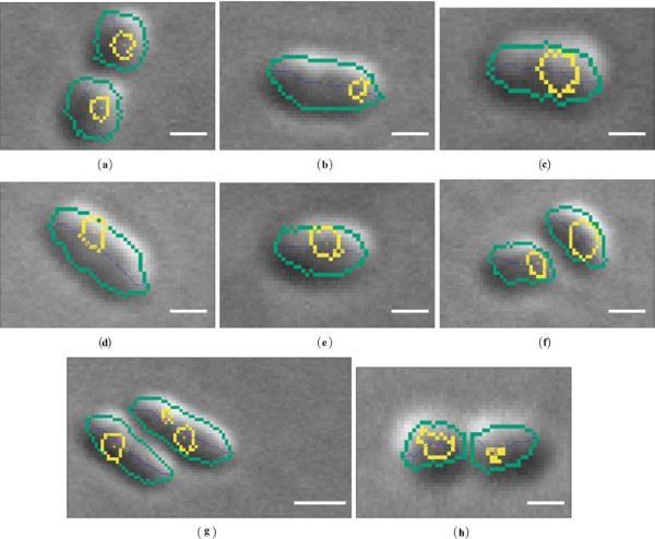
Figure 6.
Results of the proposed approach for the analysis of the spatial relations between the cell and its chemotaxis proteins (a-h). Legend: (green) - cell boundary, (yellow) - chemotaxis protein boundary, (yellow circle) - cell centroid position (r), (yellow star) - chemotaxis protein position (ci), (magenta star) - point on the centreline which correspond to ci, (green cross) - arbitrary end of the cell. Scale bars correspond to 1 [μm].

