
Figure 7.
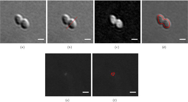
Bacterial cell and chemotaxis protein body segmentation. Bacterial cell and chemotaxis protein body segmentation workflow: (a) DIC input image, (b) DIC shear direction estimation: an obtained orientation aμ = 48.20 [deg] (red line) calculated by use of a rotated linear structuring defined by l = 20 [pixels] and n = 180, (c) DIC image reconstruction, (d) an overlay of the segmented cell body in red, (e) fluorescent input image, (f) an overlay of the segmented chemotaxis protein body in red. Scale bars correspond to 1 [μm].