
Figure 8. SporoAMA1 DIII reorganization upon ligand binding provides possible insight into signal transduction mechanisms.
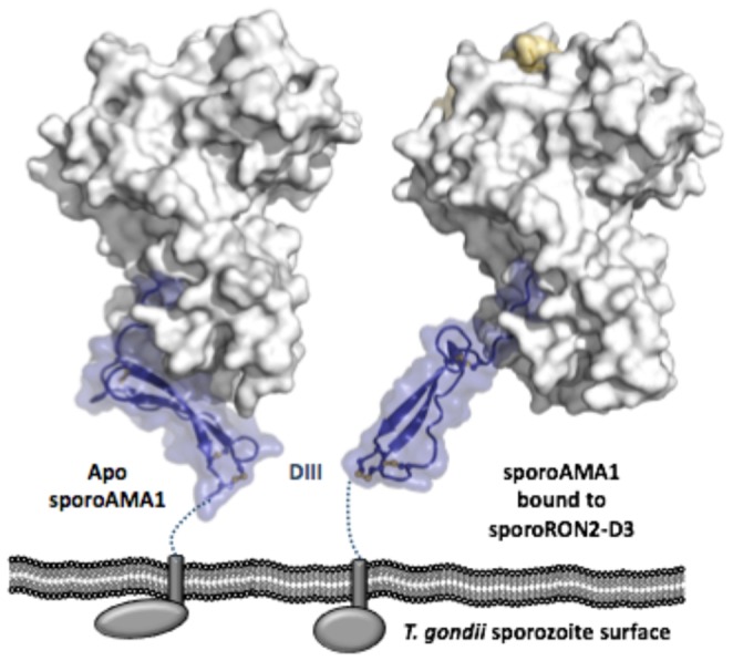
SporoAMA1 shown in predicted organization to the T. gondii sporozoite plasma membrane, with DI and DII shown as a grey surface and DIII shown as blue cartoon with a semi-transparent blue surface, sporoRON2-D3 shown as a gold surface, and disulfides shown as yellow sticks. Dotted lines indicate extended Pro/Glu rich region between the conserved portion of DIII and the transmembrane domain (grey rectangle) that leads through to the C-terminal domain (grey oval/sphere). Left – Apo SporoAMA1. Right – SporoAMA1 bound to sporoRON2-D3.