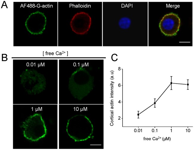
Figure 4. Calcium-dependent cortical actin polymerization in permeabilized chromaffin cells.
Cultured chromaffin cells were permeabilized in buffer KGEP (mM: 139 K+-glutamate, 20 Pipes, 5 EGTA, 2 ATP-Mg and 0.01 free calcium, pH 6.6) during 6 minutes with 20 µM digitonin in the presence of 0.3 µM Alexa-Fluor488-G-actin conjugate (AF488-G-actin), fixed and visualized by confocal microscopy. A: Total F-actin was stained using 1 µM phalloidin-rodhamine B (red) and nuclei were stained with 5 µg/ml DAPI (blue). Note that newly synthesized actin was incorporated into pre-existing cortical filaments. B–C: The new formation of cortical actin filaments was assessed by quantifying AF488-G-actin staining mean intensity at the cell periphery in the presence of increasing free Ca2+ concentrations. Note that maximal cortical actin polymerization was observed at a range of 1–10 µM of free Ca2+. Scale = 10 µm. Data are means of cortical actin fluorescence intensity from at least 12 cells per each Ca2+ concentration (12 cells for 0.01 µM Ca2+, 13 cells for 0.1 µM Ca2+, 15 cells for 1 µM Ca2+,and 18 cells for 10 µM Ca2+).

